Abstract
Introduction: Neuroendocrine carcinoma coexisted with adenocarcinoma in cervix is very rare. This disease have a very poor prognosis and so far no standard treatment has been obtained.
Case Illustration: A 39-years-old woman seeks medical attention with chief complaint of bleeding outside of menstruation. Ultrasound: EL (+) enlarged portio with a mass of 3.8 cm × 2.7 cm. Microscopic biopsy conclusion was obtained a mixture of adenocarcinoma and neuroendocrine cervical carcinoma, moderate-bad differentiation. Several Immunohistochemical staining of the biopsy tissue was done to detect neuroendocrine and adenocarcinoma. Also P63 was performed to dismissed squamous cell carcinoma.
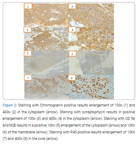
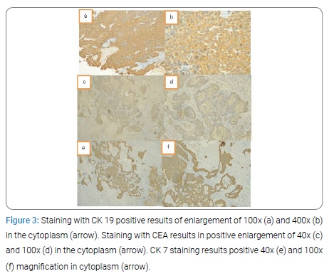
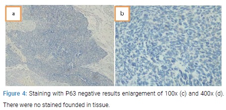
Discussion: In biopsy tissue, P63 immunohistochemical staining was performed to detect squamous cell carcinoma with negative results. Furthermore chromogranin, synaptophycin, CD 56, NSE, and P40 immunohistochemical staining were performed to detect neuroendocrine with positive results and so did CK 19, CEA, and CK 7 staining to detect positive adenocarcinomas.
Conclusion: This rare case had a positive immunohistochemical staining of both neuroendocrine carcinoma and adenocarcinoma cervix.
Introduction
A mixture of adenocarcinoma and neuroendocrine carcinoma in the cervix is atumor that have characteristics of neuroendocrine differentiation and cervical adenocarcinoma, this disease is very rare. Neuroendocrine tumors in the cervix can arise from neuroendocrine granule cells that are normally present in the cervix or from the cervical reserve/stem cells. There are 4 classifications of this type of tumor, namely typical carcinoid tumors, atypical carcinoid tumors, small cell carcinomas, and large cell carcinomas [1]. Small cell carcinoma types are more common than other types, which is around 2% to 5% of all cervical cancers [2]. Some studies state that only 4% of small cell carcinomas are coexistent with adenocarcinoma [3]. This type of tumor has a poor prognosis and so far no standard treatment has been obtained for this type of tumor [4].
Case Presentation
A 39-years-old woman seeks medical attention with chief complaint of bleeding outside of menstruation. This bleeding persisted as from the last year accompanied by discomfort in the lower abdomen. Patients also experience vaginal discharge accompanied by blood since the last 6 months. Patient’s history of using 3-month injection hormonal contraception. The physical examination showed an exophytic mass of 5 cm diameter in the portio of cervix. Ultrasound: EL (+) enlarged portio with a mass of 3.8 cm × 2.7 cm. Temporary diagnosis of this patient is cervical Ca stage II and recommended for biopsy of the cervix.
Microscopic biopsy results: Invasive epithelial malignant tumorinto the connective tissue formed mostly solid structures, sheets, papillaries, and few form glands and cribiform structures (Figure 1).
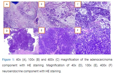
Tumor cells were round, pleiomorphic, hyperchromatic, partly with a molded core, granular chromatin/partly vesicular with a real child nucleus. Eucinophilic cytoplasm.Also visible extracellular mucin. Mitosis found. Conclusion was obtained a mixture of adenocarcinoma and neuroendocrine cervical carcinoma, moderate-bad differentiation. Immunohistochemical staining of the biopsy tissue was done by staining Chromogranin, synaptofisin, CD 56, NSE, and P40 to detect neuroendocrine (Figure 2).
P63, CK 19, CEA, and CK 7 staining was also performed to detect adenocarcinoma and P63 to detect squamous cell carcinoma (Figure 3,4).
Discussion
I report a very rare case, a mixture of neuroendocrine carcinoma and adenocarcinoma of the cervix. Patients have clinical manifestations of bleeding outside the menstrual cycle accompanied by vaginal discharge and discomfort in the hip. This is consistent with the theory that states that tumors with small and large cell neuroendocrine carcinoma components have manifestation of vaginal bleeding, cervical mass, and/or abnormal cervical cytology. In biopsy tissue, P63 immunohistochemical staining was performed to detect squamous cell carcinoma with negative results. This was done to eliminate the diagnose of squamous cell carcinoma. Usually, more frequentcases founded were squamous cell carcinoma or a mixture of squamous cell carcinoma with adenocarcinoma (adenosquamos carcinoma). Furthermore chromogranin, synaptophycin, CD 56, NSE, and P40 immunohistochemical staining were performed to detect neuroendocrine with positive results and so did CK 19, CEA, and CK 7 staining to detect positive adenocarcinomas. This shows that this tissue is indeed a mixed case between neuroendocrine carcinoma and adenocarcinoma in servix. In biopsy tissue, a mixture of neuroendocrine carcinoma and adenocarcinoma is suspected,immunohistochemical staining should also be done to detect adenosquamouscarcinoma. So that patients could get appropriate management and prognosis.
Conclusion
This case is a mixture of neuroendocrine carcinoma and adenocarcinoma in servix due to the positive markers supporting the carcinoma.
Keywords
Neuroendocrine carcinoma; Adenocarcinoma cervix; Immunohistochemical
Cite this article
Primariadewi Rustamadji. Adenocarcinoma admixed neuroendocrine carcinoma: a rare case report of 39 years old women. Clin Oncol J. 2020;1(1):1–3.
Copyright
© 2020 Primariadewi Rustamadji. This is an open access article distributed under the terms of the Creative Commons Attribution 4.0 International License (CC BY-4.0).